To fully understand how a thermal image, or thermogram is produced, some background knowledge of the physics of light is needed.
Light is emitted in waves. The amount of energy in each light wave is related to its wavelength; shorter wavelengths have more energy.
Visible light is made up of a spectrum of colours (those you seen in a rainbow), in this spectrum violet has the most energy and red has the least.
As well as the visible light spectrum there are also UltraViolet (UV) and InfraRed (IR) Spectrums either side of it. These can’t be seen with the naked eye.
The IR Spectrum is the part of light that we’re interested in when capturing IR images.
Infrared light can be split into three categories;
1. Near IR; closest to visible light, near infrared has wavelengths ranging from 0.7 to 1.3 microns (or 700 to 1300 billionths of a metre);
2. Mid-IR; has wavelengths ranging from 1.3 to 3 microns;
3. Thermal-IR; has wavelengths ranging from 3 to 30 microns.
You’re probably very familiar with both near and mid-InfraRed as they’re used by a variety of electronic devices, e.g. remote controls. Thermal-IR makes up the largest part of the IR Spectrum and is also known as heat. Thermal-IR is the part of the Spectrum we’re interested in for Thermography (Thermal Imaging).
The key difference between Thermal-IR and the other categories is that it’s emitted by an object instead of being reflected off it.
Animals emit IR radiation as a result of normal physiological processes. About 60% of all the heat an animal produces passes into the surrounding air as radiant heat, or heat photons. The remaining heat is lost to the environment by evaporation (25%), conduction to objects (3%) and conduction to the air (12%).
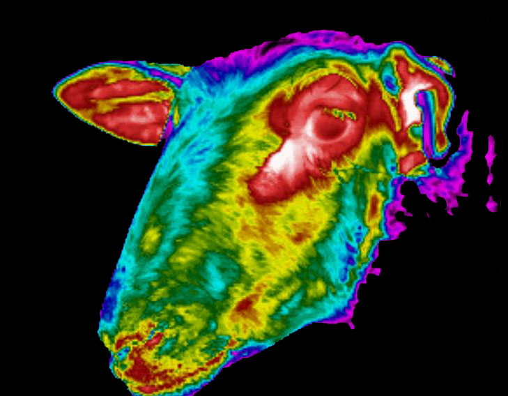
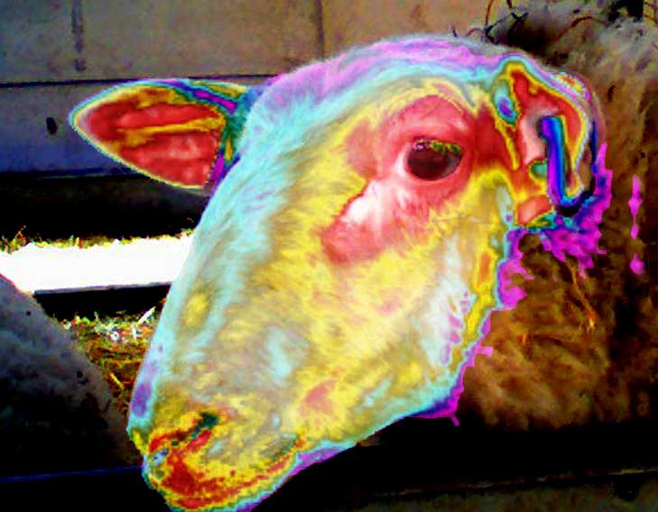
An IR camera is used to measure the heat photons emitted from the animal and to convert them into electrical impulses which are then displayed as coloured images on a monitor. This visual image graphically maps the animal’s body temperature and is referred to as a thermogram. The heat detector, or microbolometer within the thermal imaginng camera can detect differences in temperature of less than 0.05oC, which is 40 times more sensitive than the human hand. More reading about heat detectors can be found on Wikipedia.
These pictures were taken with the thermal imaging camera, which has the ability to simultaneously take and then fuse together visible light and thermal images.
Wool is a fantastic insulator, so heat is not emitted through her fleece, and this area doesn’t even register on the thermal scale. The corners of the eye are always the warmest points on any animal as the skin is thinner here, allowing heat to radiate out easily. As the ear is concave, the infrared radiation bounces around and concentrates in this area making it appear warmer.
This article comes from veterinary-thermal-imaging edit released